Mazen Ibrahim, MD
Assistant Professor, Department of Orthopaedic Surgery
SUNY Downstate Health Sciences University
Office: 718-270-4335
mazen.ibrahim@downstate.edu
Introduction
The hip joint is made up of: a ball on the upper end of the thigh bone (femur), called the head of the femur, and a socket in the pelvis known as the acetabulum. An extremely low friction tissue, hyaline cartilage, lines the joint. Normal acetabulum "covers" the upper (superior) portion of the head of the femur as well as a portion of the front (anterior) and back (posterior) of the femoral head.
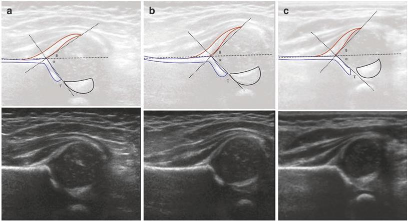
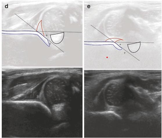
Hip Dysplasia or developmental dysplasia of the hip includes several morphologic abnormalities caused by intrauterine mechanical factors leading to dysplastic changes of the femoral and acetabular sides that continue until the bones reach maturity. Hip dysplasia is, at its core, a condition of instability. This instability can cause mechanical overloading of the acetabular rim during normal activities with associated increased stresses in the cartilage which can lead to chondral and labral damage and eventually arthritis. Acetabular dysplasia has been linked to increased intra-articular biomechanical forces leading to labral tears and articular cartilage degeneration.
Periacetabular osteotomy
Periacetabular osteotomy (PAO) represents the cornerstone in the treatment of acetabular dysplasia, with several long-term studies demonstrating clear benefits in regard to patient function, cartilage health and preventing degenerative arthritis. The goal is to alleviate the patient's pain, restore function, and maximize the functional life of their dysplastic hip. PAO is a procedure that was developed and first performed in 1984 in Bern, Switzerland by Professor Reinhold Ganz. "Periacetabular" means around the acetabulum. "Osteotomy" means to cut bone.
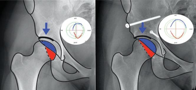
The PAO cuts the bone around the acetabulum that joins the acetabulum to the pelvis. Once the acetabulum is detached from the rest of the pelvis by a series of carefully controlled cuts, it could be rotated to a position of ideal coverage as dictated by the specific acetabulum's unique anatomy. Intraoperative X-rays is utilized to confirm the desired correction of the acetabulum is achieved and screws are inserted into the bone to maintain the acetabulum in its new corrected position during bone healing.
PAO thereby reorients the acetabulum and brings the roof from an oblique to a horizontal position. The dysplastic roof that incompletely covers the femoral head is brought over the head to give the head a normal coverage. Other subtle changes typically occur for the anterior and/or the posterior femoral head coverage.
Postoperative
The first 2-3 hours following surgery, the patient is closely monitored in the recovery room. On average, patients spend 1-2 nights in the hospital recovering, and receiving medications for pain control, preventing infection, and the prevention of deep vein thrombosis (blood clots in large veins), and pulmonary embolus (blood dots traveling through veins to the lungs). Patients begin physical therapy and occupational therapy as soon as possible, while they are in the hospital, to improve hip motion, muscle function and to learn how to use appropriate assistive devices such as crutches or walkers. X-rays will be taken 1-2 days after surgery for a final assessment of the result.
After discharge
The hospital stay is 1-2 days, depends on how rapidly pain subsides and progress in physical/occupational therapy. At discharge pain medication is prescribed as well as an anticoagulant to prevent blood clots, in addition to OTC medications for constipation, insomnia and itching as needed.
Some degree of pain after discharge is natural, varies from one patient to another, which may increase or decrease on different days, but the general trend should be toward decreasing pain. Numbness and a tingling sensation are common around the incision area.
The skin incision is closed with dissolvable sutures under the skin, surgical glue and outer water proof dressing. The outer dressing could be changed if soaked or peeled. The surgical glue shouldn’t be removed, approximately 2 weeks. The patient can shower starting the day after surgery. Water is allowed to run over the incision site, but avoiding tubs, pools or any soaking or scrubbing of the surgical site for at least two weeks. Bruising in the thigh and calf is normal with surgery and may not appear until several days afterwards. Some spotting from the incision is normal, however, if dressing soaking is significant, please notify us.
Thromboembolic Deterrent (TED) stockings, Compression Socks and Sequential Compression Device (SCD) machines will be prescribed at surgery. TED stockings should be worn for 3-4 weeks postoperatively to reduce the risk of blood clots in the legs (deep vein thrombosis/DVT). TED stockings may be removed for showering and for sleeping. In addition to the TED stockings, it is recommended to getting up and moving around every 60-90 minutes post-operatively.
During the first six weeks following surgery, the injured hip should bear no more than a limited weight of 30 pounds. Placing full weight on the operated side prior to bone healing can cause the screws to bend or break and the osteotomy to lose its position. Too vigorous exercise such as resistive exercise against weights can also cause failure. If failure occurs, re-operation may be necessary, and the chance of developing arthritis is greatly increased.
Follow-up outpatient visits are necessary to monitor progress by physical examination and X-ray. The first follow-up visit is usually scheduled about 2 weeks after surgery to check on the wound healing and to confirm if the patient no longer needs prescribed pain medications, and the second at 6 weeks after surgery. At 6 weeks after the surgery the patient is allowed to be full weight bearing and to work toward discontinuing use of the crutches. Muscle strengthening exercises often with the help of a physical therapist are also started. Progress in walking depends on the osteotomy healing on X-rays and the muscle strength recovery. Subsequent follow-up visits are at 3-months 1-year and 2 years after surgery and then at 2-3 years intervals. If the patient requested, removal of the screws that were used to fix the PAO can be performed as an outpatient procedure, that does not interrupt a patient's continued full function.
Outcome
While several surgical options have been described for the treatment of acetabular anatomic abnormalities, PAO remains the preferred option, giving marked improvements in functional outcomes as well as preventing long-term cartilage degeneration. Optimization of the joint surface area in the weightbearing region of the hip and improvements in the mechanical advantage of the abductor musculature are thought to provide most of the symptomatic improvements postoperatively. PAO outcomes have shown good symptomatic improvements, low complication rates, and an 18-year hip joint survival rate of >75%.
In the past decade, a strong emphasis has been placed on patient-reported outcome measures (PROMs) to better define quality metrics guiding the clinician on how to best optimize the clinical outcomes of patients suffering from symptomatic hip dysplasia undergoing PAO.
References
- Ibrahim, M. M., Smit, K., Poitras, S., Grammatopoulos, G., & Beaulé, P. E. (2021). Correlation of Patient-Reported Outcomes After Periacetabular Osteotomy with Femoral Head Coverage and Acetabular Orientation: A Single-Center Cohort Study. The American journal of sports medicine, 49(5), 1209–1219.
- Ibrahim, M. M., Poitras, S., Bunting, A. C., Sandoval, E., & Beaulé, P. E. (2018). Does acetabular coverage influence the clinical outcome of arthroscopically treated cam-type femoroacetabular impingement (FAI)? The bone & joint journal, 100-B(7), 831–838.
- Ibrahim M. M., Smit K. (2020) Anatomical Description and Classification of Hip Dysplasia. In: Beaulé P. (eds) Hip Dysplasia. Springer, Cham
- Wilkin, G. P., Ibrahim, M. M., Smit, K. M., & Beaulé, P. E. (2017). A Contemporary Definition of Hip Dysplasia and Structural Instability: Toward a Comprehensive Classification for Acetabular Dysplasia. The Journal of arthroplasty, 32(9S), S20–S27.
- Bali, K., Smit, K., Ibrahim, M. M., Poitras, S., Wilkin, G., Galmiche, R., Belzile, E., & Beaulé, P. E. (2020). Ottawa classification for symptomatic acetabular dysplasia assessment of interobserver and intraobserver reliability. Bone & joint research, 9(5), 242–249.
- Ziran, N., Varcadipane, J., Kadri, O., Ussef, N., Kanim, L., Foster, A., & Matta, J. (2019). Ten- and 20-year Survivorship of the Hip After Periacetabular Osteotomy for Acetabular Dysplasia. The Journal of the American Academy of Orthopaedic Surgeons, 27(7), 247–255.
University Orthopaedic Associates
Department of Orthopaedic Surgery & Rehabilitation Medicine
DOWNSTATE HEALTH SCIENCES UNIVERSITY
450 Clarkson Avenue, Box 30, Brooklyn, NY 11203 · Phone 718-270-2045